Nervous system or nervous
co-ordination
The system which co-ordinate, regulate and controlled all
other systems is called nervous system. Functional Unit of nervous system is neuron
or nerve cell. Nervous system of higher animal performs three
functions these are Receiving, Processing or Modulating and
Responding.
Function of Nervous system.
- The stimuli or impulses are carried to CNS from muscles and vice versa.
- Co-ordinates various organs system.
- It stimulates and inhibits the activities of muscles, glands and viscera.
- It helps to maintain the homeostatic condition.
Parts of nervous system
- Central nervous system (CNS)-It includes brain and spinal cord.
- Peripheral nervous system (PNS)-It includes the nerves arising from CNS.
- Autonomic nervous system (ANS)-It includes nerves and ganglia extending up to visceral organs. It works involuntarily.
- Central nervous system
CNS consist of two parts-
1. Brain
Brain is highly specialized delicate organ located in skull
or cranium. It is about 1.4 kg. Brain is covered by three layers known as meninges.
I. Outer
layer is duramater-It is just located below the skull. It is highly
vascular tough white fibrous tissue. It supports the brain spinal cord.
II. Middle
layer is arachnoid-It is the middle, delicate and thin fibrous covering
below the subdural cavity.
III. Inner
layer piamater-It is thin delicate and highly vascular innermost layer.
It is separated from arachnoid membrane by a subarachnoid space. The
space is filled with spongy connective tissue and cerebrospinal fluid (CSF).
Cerebrospinal fluid (CSF)
It is secreted from anterior and posterior choroids plexus.
It performs following functions.
- Act as cushion shock absorber.
- Act as medium for exchange of different materials through the blood vessels.
- Keep the brain and spinal cord moist.
- It makes the brain weight less.
Structure of human brain
Human brain is divided into three parts;
1.
Fore brain(prosencephalon)
2.
Mid brain(Mesocephalon)
3.
Hind-brain(Rhombencephalon)
 |
Fig: mid-sagittal
section of human brain
|
Forebrain is distinguished in to 3 parts-
- Olfactory lobes
- Cerebrum
- Diencephalon
- Olfactory lobes have anterior olfactory bulb and posterior olfactory tract. These parts are fully covered by cerebral hemisphere and ventral view can be seen. Cavity of lobes is called 1st ventricle. Olfactory lobes are poorly developed in human. Function of olfactory is to receive the sense of smell.
Fig: Lobes of cerebrum

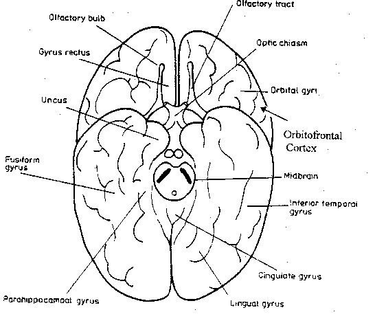
fig: Olfactory lobes
- Cerebrum is more complex and large part of brain. It is divided in to left and right hemisphere. Both hemispheres are connected by nerve fibers called corpus callosum. Outer layer of cerebrum is known as cerebral cortex formed of convolutions. Raised parts are called gyri and depressions are called sulci. Each hemispheres can be divided in to- frontal parietal, temporal and occipital lobes by sulci.
There are different areas on the cerebrum these are-Motor
area for movement, sensory area for heat, cold, light,
pressure, touch etc auditory area for hearing, visual area for
seeing olfactory area for taste and smell, taste area and smell
area. Each hemisphere receives information from opposite side of the body.
Cavity of each cerebral hemis- sphere is called lateral ventricles.
Functions-
a)
It controls all mental and conscious activities (such as intelligence,
memory, reason, will, feelings and emotions).
b)
It is the site of originator of voluntary acts and interpreter of
sensations.
c)
It is a control center of reflex actions.
3. Diencephalon lies just below the corpus
callosum and the mid brain. It has three parts-epithalamus,
thalamus and hypothalamus. Epithalamus is anterior part and folded
to form anterior choroids plexus which secrete CSF. There is a pineal body behind
the choroids plexus. It secrets melatonin hormone.
Thalamus is the part which consists of optic
chaisma (crossing of optic nerves). It is a sensory relay station.
Hypothalamus consist of a hypophysis (pituitary
gland) attached by stalk called infundibulum. Pituitary gland
is a master gland. The cavity behind the lateral ventricle is
called third ventricle. It is connected with ventricle of cerebrum by foramen
of monro.
Functions:
a)
It serves as relay center between spinal cord and brain stem.
b)
It regulates emotions and perceptions.
Mid-brain
It is the brain which connects
forebrain with hind brain. It has two parts;
Corpora quadrigeminia; These are two pairs of round
lobes.1stpair is called superior colliculi which concern with
vision.2nd pair is called inferior colliculi which concern
with hearing.
Crura cerebri; These are two bundles of fibers which
lie on the lower surface of the mid brain. It relay the impulses back and forth
between the cerebrum, cerebellum, pons and medulla oblongata.

Hind brain
It consists of cerebellum, pons varolli and medulla
oblongata.
Cerebellum; It is the second largest part of brain
located behind posterior part of brain. It has three lobes. Cavity of
cerebellum is called fourth ventricle communicating with third ventricle
through aqueduct of sylvius.
Functions
a)
Co-ordinate muscular body movement.
b)
It controls reflex action of skeletal muscle activities.
Pons varollii (pons =bridge); it is situated in front
of cerebellum and above the medulla oblongata and join medulla oblongata with
the mid brain. Its fibre is of white matter.
Medulla oblongata; It lies between the pons varolii
and the spinal cord. It consists of white matter. It is continuous with the
spinal cord. Lower part of it consists of non vascular folded structure called posterior
choroid plexus.
Functions;
a)
It is centre for cardiac, respiratory and vasomotor.
b)
It controls complex activities such heart action, respiration, sneezing,
coughing etc.
Q. Write differences between cerebrum and cerebellum.
Brain Stem
Mid brain, pons and medulla oblongata collectively forms the
brain stem. It connects the fore brain with spinal cord. It is continues with
spinal cord. Death is declared clinically when there is cessation of brain stem
function.
Ventricles of Human Brain
Ventricles are the cavities within the brain filled with
cerebrospinal fluid (CSF).It secreted by anterior and posterior plexuses. There
are four ventricles.
a)
Right and left lateral ventricles.
b)
Third ventricle (Dioecoel)
c)
Fourth ventricle (Metacoel)
Right and left lateral ventricles lie within the
cerebral hemispheres below the corpus callosum. They communicate with third
ventricles by foramen of monro.
Third ventricle situated below the lateral ventricles
between two parts of the thalamus. It is communicated with third ventricle by
narrow canal iter or cerebral aqueduct.
Fourth ventricle is a lozenge-shaped cavity situated
below and behind the third ventricle, between the cerebrum and pons varolii. It
continues below the central canal of spinal cord.

fig: ventricles of brain
Cerebro-Spinal Fluid (CFS); It is lymph like
intracellular fluid. It consists of salts, glucose, and small amount of
albumin, globulin and few traces of urea and creatinine.
CSF is secreted by anterior and posterior choroid
plexus in ventricles of brain, central canal or spinal cord and apace around
the brain and spinal chord. Total volume of CSF is 150ml.the rate of formation
is 20ml/hr.CFS can be obtained by lumber puncture.
Function of CSF
- It protects brain and spinal cord.
- It maintains uniform pressure around these delicate structures.
- It acts as a cushion and shock absorber.
- It keeps the brain and spinal cord moist and exchange of substances between CFS and nerve cells.
- It excretes harmful metabolic wastes, drugs and other substances from the brain to the blood.
- It nourishes the nerve cells.
- It also acts as buffer.
- It act as buoyancy and makes the brain low weight.
2. Spinal cord
It is a posterior part of CNS which runs mid dorsally within the vertebral
column. It is elongated, almost cylindrical part. It extends from the medulla
oblongata within vertebral column to the level of second lumber vertebra. It
measures about 42 to 45cm long and 2 cm thick. It is surrounded by the same
three meninges found in the brain.
Internal structure
Internally, spinal cord is divided in to left and right symmetrical halves.
Posterior is median sulcus and anterior is median fissure. In the there is a
central canal surrounded by a butterfly shaped area of grey matter. Around the
grey matter there is white matter. Grey matter is an H-shaped with two dorsal
and ventral horns. Roots of spinal nerve are originated from the horns. There
are 31 pairs of spinal nerves arise from different segments of spinal cords.
Each spinal nerve carries both sensory and motor impulses. Each spinal nerve
connects with nerve roots.
/17%20Control%20and%20Coordination/img%201/3.jpg)
Fig:
T.S. of spinal cord
- Dorsal nerve root: It originates from the dorsal horn of grey matter. It consists of only sensory fibers. It bears the dorsal root ganglion containing only sensory cells.
- Ventral nerve root: It originates from the ventral horn of grey matter. It is made up of only motor fibers. It does not bear ganglion.
Functions of spinal cord
i.
It is center of spinal reflex action.
ii.
The stimuli are passed from and to the brain through the spinal cord.
Q. Write differences between ascending tract and
descending tract.
Q. Write differences between grey matter and white matter
of CNS.
- Peripheral Nervous System(PNS)
It consists of:
- Cranial nerves: Those arising from the brain.
- Spinal nerves: those arising from the spinal cord.
Types of Nerve Fibres
1. Types on the basis of
structures:
a.
Myelinated or medullated nerve fibres-
These fibre have myelin sheath. These are found in white
matter of brain, spinal cord and nerves.
b.
Non-myelinated or non- modulated nerve fibre-These are without myelin
sheath. They are found in grey matter of brain and spinal cord.
2. Types on the basis of function
of nerve impulse:
a.
Afferent nerve fibres (sensory): These conduct nerve impulses from
effectors organs to the CNS. Such as optic nerve.
b.
Efferent nerve fibres (motor): These conduct nerve impulses from CNS to
body organs. Such as occulomotor nerve (for eye movement).
c.
Mixed nerve fibres: These are both sensory and motor in function such as
spinal nerves.
3. Types on the basis of number of
their processes
a.
Unipolar: These neurons have only one axon with dendrites. They
are found in dorsal root ganglia of spinal nerve.
b.
Bipolar: These neurons have two processes. In which one may be
dendrite and other is axon. These are found in the retina of eye.
c.
Multipolar: These neurons have many cell processes. These are
found in CNS.
Cranial nerves
There are 12 pairs of cranial nerves. These arise
from ventral side of brain. There names types and functions are given below in
table.
No.
|
Cranial nerve
|
Types of fibre
|
Organs innervated
|
Function
|
I
|
Olfactory nerve
|
Sensory
|
Mucosa in nose
|
Smell
|
II
|
Optic nerve
|
Sensory
|
Retina of eye
|
Vision
|
III
|
Occulomoror nerve
|
Motor
|
Eye muscles, Ciliary muscles
|
Eye movement, accommodation
|
IV
|
Trochlear nerve
|
Motor
|
Superior oblique muscles of eye ball
|
Eye movement
|
V
|
Trigeminal nerve
|
Mixed
|
Skin teeth, mucosal membrane of mouth
|
Sensation head face
|
VI
|
Abducens nerve
|
Motor
|
Eyeball muscles
|
Eyeball movement
|
VII
|
Facial nerve
|
Mixed
|
Taste buds, salivary glands, facial and neck muscles
|
Facial expression, saliva secretion, taste
|
VIII
|
Auditory nerve
|
Sensory
|
Internal ear
|
Equilibrium Hearing
|
IX
|
Glossopharyngeal
|
Mixed
|
Pharynx, tongue, salivary glands
|
Taste, swallowing and saliva secretions
|
X
|
Vagus nerve
|
Mixed
|
Pharynx to viscera
|
Visceral reflexes
|
XI
|
Spinal accessory
|
Motor
|
Thoracic and abdominal viscera
|
Visceral reflexes, Shoulder movement
|
XII
|
Hypoglossal nerve
|
Motor
|
Muscles of tongue
|
Movement
|
Spinal Nerves
There are 31 pairs of spinal nerves in human immersed from
either side of spinal cord from intervertebral foramina.
These nerves are:
Cervical
8 pair in neck
Thoracic
12 pair in thorax
Lumber
5 pair in upper abdomen
Sacral
5pair in lower abdomen
Coccygeal
1 pair in tail region
Total spinal fibers
= 31pairs
So the spinal formula is --- C8
Th12 L5 S5 Co1
Each spinal nerve is a mixed nerve. It originates by two
roots-Dorsal or sensory or afferent roots and Ventral or
motor or efferent root from spinal cord. The two roots join
within the neural canal of vertebral column. Each spinal nerve immediately
divides into three branches:
· Ramus
dorsalis: It supplies muscles and of dorsal side.
· Ramus
ventralis: It supplies muscles and skin of ventral and lateral sides.
· Ramus
comminicans: it joins sympathetic ganglion of ANS.
Q. Write difference between cranial nerves and spinal
nerves.
Autonomic Nervous Sys tem (ANS)
Autonomic nervous system controls and co-ordinates the
various activities of visceral organs. Hence it is also called visceral
nervous system. Actually ANS is not autonomous or independent
because it is regulated by higher nerve centre of brain. It consists of two
antagonistic (opposite in function) systems-
1. Sympathetic nervous system
2. Parasympathetic nervous system
Sympathetic nervous system
It consists of sympathetic chains, preganglionic
fibres, collateral ganglia and postganglionic sympathetic fibres.
i.
Sympathetic chains: There are long lateral chains of sympathetic
ganglia (21=3 cervical, 12thoracic, 5 lumbar and 1 sacral) present on
either side of vertebral column.
ii.
Preganglionic sympathetic fibres: There are short sized axons
originate from the grey matter of spinal cord.
iii.
Collateral ganglia: There are three collateral ganglia-coeliac
ganglion, superior mesenteric ganglion and inferior mesenteric ganglion.
iv.
Postganglionic sympathetic fibres: These are long sized axon of
neurons originated from collateral ganglia. These nerves supply the
visceral organs and iris and ciliary muscles.
The sympathetic nerves stimulate the adrenal glands to
secrete adrenalin or nor adrenaline, so these are called adrenergic
nerve fibres.
Diagram……
Parasympathetic nervous system
It consists of parasympathetic fibres, parasympathetic
ganglia and postganglionic parasympathetic fibres.
i.
Preganglionic parasympathetic fibres: These are long sized axons
of neurons present in midbrain brain stem and sacral region of spinal cord.
These are emerging from cranium and sacrum form cranio-sacral out flow.
ii.
Parasympathetic ganglia: These ganglia are present either close
or inside the muscles of visceral organs. These are isolated ganglia. These
ganglia join the preganglionic fibres and post-ganglionic fibres.
iii.
Postganglionic parasympathetic fibres: These are short sized
axons of neurons arising from the parasympathetic ganglia and supply smooth
muscle and glands or visceral organs.
Diagram……
Q. Write differences between sympathetic and
parasympathetic nervous system.
Synapse
Synapse is an area of functional contact between one neuron
and another to transfer information. Synapses are usually found between the
fine terminal branches of the axon of a neuron and the dendrites or cell body
of another.
Diagram….
Structure of synapse
A typical synapse consists of a bulbous expansion of a nerve
terminal called a pre-synaptic knob close to the membrane of a dendrite.
Cytoplasm of synaptic knob contains mitochondria, smooth endoplasmic
reticulum, micro-filaments and numerous synaptic vesicles. Each vesicle
contains neurotransmitter responsible for transmission of nerve impulse across
the synapse. The membrane of synaptic knob nearest the synapse is thickened and
forms the pre synaptic membrane. These membranes are separated by a gap, the synaptic
cleft. The post-synaptic membrane contains large protein molecules which
act as receptor sites for neurotransmitter and numerous channels and pores.
The two neruro-transmitters in vertebrate nervous are
acetacholine (Ach) and noradrenalin although other neurotransmitters
also exist. Neurons which release the neurotransmitters are called cholinergic
neurons and adrenergic neurons.
Diagram….
Stimulus
Stimulus is a sudden change in the external or internal
environment, which excite the nerve or organism or muscle as whole. The
stimulus which capable to just excite given tissue is called threshold
stimulus.
There are many types of stimuli which can excite the tissue:
a.
Mechanical stimuli- These include touch muscular stress etc.
b.
Physical stimuli-These include heat and humidity.
c.
Chemical stimuli-The electrical stimuli is able to excite tissue.
Properties of a nerve fibre are:
i.
Excitability
ii. Conductivity
iii. Refractory period
iv. Summation v. All or none rule.
i.
Excitability-When a nerve fibre is stimulated by stimuli of
physical, mechanical or chemical means and impulse is formed, it is called
excitability.
ii. Conductivity-When the
stimulated nerve transmits the impulse in a particular
direction is called conductivity.
iii. Refractory period-After excitation
or transmission of nerve impulse nerve
regain the original state is called refractory period.
iv. Summation-When stimulus applied to a
nerve fibre is below the threshold
stimulus then it fails to stimulate any response. However, if the
same
stimulus is continuously applied, stimulation occurs. This is called summation.
v. All or none rule-When the organism
gives response by stimulation, it cannot be increased or
response is always maximum. It is called all or none rule.
Nerve impulse
A nerve impulse is defined as wave of depolarization (wave
of reverse polarity) of the membrane an axon of nerve cell. Nerve impulse is
generated in nerve fibre is an electrical phenomenon.
Extra cellular fluid and intracellular fluid
Nerve is bathed in side the fluid. Outside the nerve is
extracellular fluid (ECF) and inside the nerve fibre is the intracellular fluid
(ICF).
· Extra
cellular fluid contains a large amount of sodium chloride, bicarbonates,
oxygen, carbon dioxide and other metabolic wastes.
· Intracellular
fluid contains a large amount of potassium and magnesium phosphates along
with proteins and organic molecules.
Most of the solutes in
extracellular fluid and intracellular fluid are electrically charged particles
or ions (K+, Na+).
Differential
permeability: Extra cellular fluid contains ten times more Na+
ions than inside the membrane of nerve cell which make outer side
electropositive. K+ ions are 25 times more in intracellular fluid
which makes inner side membrane electronegative. It is due to the
differentially permeable to Na+, K+, Ca++ , and
Cl- ions. Cell membrane of
neuron is less permeable for Na+ but it is more permeable for K+
(about 50 times more than Na+).this phenomenon is called
differential permeability. It is maintained by Na-k pump.
Transmission of nerve
impulse
It is described in two steps.
A. Transmission of
nerve impulse along the nerve fibre.
B. Transmission of
nerve impulse across the synapse.
Transmission of nerve impulse along the nerve fibre is
explained by Hodgkin and Huxley in the late 1930 in three steps.
- Polarization or resting potential.
In the rest state the inside of
membrane has –ve electrical potential compared to outside.
This difference in potential is
called resting potential. Which is about -40mv to-90mv.Na+ and K+
are transported across the membrane against their concentration gradient by
carrier protein. Which is called Na-k pump and energy is used through ATP. The
sodium channel and potassium channels on the membrane of neuron are closed.
- Depolarization or action potential.
A nerve impulse can be initiated
by mechanical, chemical and physical stimulation. Sodium channels are opened
but potassium channels are closed and Na+ ions flood in through cell
membrane and create a positive charge of +40mv. It is very short periods that
change in potential and last for 3 milliseconds. When an action potential
occurs, the axon is said to be depolarized.
Diagram(consult transmission of nerve impulse)
- Repolarization.
Sodium channels are closed
potassium channel are opened and K+ ions diffuse out along their concentration
gradient. This start repolarization and resting potential going to reestablish.
At the same time nerve become less permeable for Na+ than K+ .So
many K+ flow out and inside charge become more negative
than that it was originally. Na-k pump starts and normal concentrations of Na
and K ions are reestablished. Each pump actively transports two K+
ions into the cell to every three Na+ ions transported out. The
membrane is once again at its resting potential.
Transmission of nerve impulse
along a non medullated nerve fibre is slower than in medullated or myelinated
nerve fibre.
During this conduction of nerve
impulse negative charge present on outside of a depolarized area attracts the
+ve charge from outer surface of next polarized area. While +ve charge present
on inner surface of depolarized area is attracted by –ve charge on inner
surface of next polarized area.so that depolarized area become repolarized and
nest polarized area become depolarized. Hence the impulses move towards
synapse.
Transmission of nerve impulse
along medullated nerve fibre is 20 times faster than non medullated fibre. The
ionic exchange or depolarization occurs only at nodes because medullary sheath
is impermeable to ions. The action potential is conducted from node to node in
a jumping manner. This also called saltatory conduction of nerve impulses.
B. Mechanism of transmission of nerve impulse across the synapse.
It is explained by henry in 1936.Following
are the steps for the process:
- When an impulse arrives at the pre-synaptic knob of axon Ca++ ions concentrate at the synapse.
- Ca++ions pass from the synaptic cleft into synaptic knob and cause the movement of synaptic vesicles towards to the surface of knob. Vesicles discharge their neurotransmitters chemicals acetylcholine in to the synaptic cleft and return to the cytoplasm of synaptic knob to refill neurotransmitters.
- The neurotransmitter binds with protein receptor molecules in synaptic cleft. Na+ ions enter into the cell of another neuron and action potential generates on it. Thus nerve impulse transferred to the next synapse.
- The acetylcholine is hydrolyzed by an enzyme acetyl cholinesterase into acetic acid and choline in the cleft which are reabsorbed into synaptic knob and resynthesized into acetylcholine using energy from ATP.
 |
| FIG:mechanism of synapses |
Are you looking for the best treatment for parkinson's. We are here for you. We have many good experienced doctor who will treat you with there responsibility. Parkinson's treatment
ReplyDelete