CARDIOVASCULAR SYSTEM
Introduction
Blood vascular system is closed type and double circulation.
It consist of; A. heart
B. blood
vessels
C. blood
Blood vascular system balance between chemical and physical
state of body fluid. The function is called homeostasis.
A. Heart
Structure
Human
heart is myogenic (i.e. wave of
muscular contraction start from the pacemaker or AV node opposite to neurogenic). It is triangular organ,
situated between the lungs in thoracic cavity. It measures 25cm long and 250 gm
in weight. It is made up of the three layers viz; pericardium, myocardium and
endocardium. Pericardium consists of two sacs. The outer sac is made up of
fibrous tissue and the inner sac consists of double layer of serous membrane.
Pericardial cavity is filled with pericardial fluid to protect the heart from
mechanical friction and external shock. Myocardium composes cardiac muscles.
The endocardium lines myocardium and forms the valves of the heart.
Human
heart is four chambered consisting of two upper thin walled auricles and two
lower thick ventricles. The two auricles are separated each other by
interauricular septum. The right auricle is larger than the left one. The right
ventricle is larger than the left ventricle. Both ventricles are separated by
interventricular septum. Both the ventricles are thick walled. The right
ventricle is much thicker than the left ventricle. Internally both ventricles
are provided with ridges called Columnae carnae.
Auricles
open into the ventricles on the respective side by atrio ventricular apertures
which are guarded by valves. Valves check the back flow of blood. The valves
are connected with the wall of ventricles by chordae tendenae. The tricuspid
valves guard the right atrioventricular aperture and bicuspid or mitral valves
guard the left atrioventriclar aperture.
Sinu-auriclar node or pace maker situates on the wall of
right auricle near the openings of venacavas.
There
are three openings in the right auricle to collect venous blood. These are; i)
a common opening of right and left venacava (precaval vein) ii) an opening of
inferior venacava or post caval vein. iii) An opening of coronary sinuses in
posterior part of right atrium.
There
are four openings of pulmonary veins in the left auricle (2 from left lungs and
two from right lung) to collect oxygenated blood.
The
right ventricle opens into the pulmonary artery and the left ventricle opens in
to the systemic aorta. The openings of these arteries are guarded by three
semilunar valves.

Double circulation in man
There is no mixing of oxygenated and deoxygenated blood in human
body. Because of double circulation that shows.
- pulmonary circulation
- systematic circulation
Pulmonary circulation
|
Systematic
circulation
|
|
2.Significance
3. Diastole of auricles
|
The right ventricle pumps venous blood to the lungs via
pulmonary arteries.
As the blood reach the alveolar surface , carboxy
haemoglobin release carbon dioxide and receive oxygen to be oxygenated
Oxygenated blood from the lungs is carried into the left
auricles via pulmonary veins
|
The left ventricle pumps oxygenated blood to the bidy
parts via systematic aorta and arterial system.
As the blood reach to tissue level of body parts the oxyhaemoglobin
release oxygen so that oxidation of food takes place to produce energy.
Carbon dioxide produce as a by product form (bend with hemoglobin). In this
way the blood becomes deoxygenated.
The venous blood from the parts is carried into the right auricle
via two precaval and one post caval and venous system.
|
In this way two complete
circulations take place simultaneously. Therefore there is no mixing of venous
and oxygenated blood. Complete separation of auricles and ventricles and close
type of blood vascular system also forbid mixing of oxygenated and deoxygenated
blood. Coronary artery supplies the blood to heart from aorta. Coronary sinuses collect the venous blood from heart.

Origin and conduction of heart beat
Sinu-auriclar node (SA-node) or
pacemaker*, which is small pea sized structure made up of specialized
tissue situated the inner wall of right auricle near the opening of superior
venacava initiated heart beat. Therefore human heart is myogenic. The cardiac
impulse produced by the SA-node is radiated out in the form of wave not only
contract the auricles it is also be passed to the muscles of ventricular wall
due separation of auricles and ventricles by thin layer of fat i.e. annular
pad.
The AV- node is the pacemaker like
structure situated near the opening of the auriculoventricular septum. The impulses
produced by pace maker reach to AV node 0.03sec so that it can generate its waves
which will be transmitted to the myocardium viz AV bundle or bundle of His and
network of Perkinje,s fiber. The AV
bundle arises from the AV-node, descends in the interventricular septum after
crossing the fibrous ring it divides into the right and left branches. Each
branch supplies a network of Purkinje fibers into the myocardium to convey
electrical waves from AV nodes to the muscular apex so that ventricle or
contraction is possible.
 |
| FIG: Conduction of heart beat showing SA node and AV node |
Heart beat
The rhythmic contraction and
relaxation of cardiac muscle in the heart is known as heart beat. One heart
beat includes atrial systole, joint diastole and ventricular systole.
Cardiac Cycle
Atrial systole
Both the auricles contract
simultaneously due to initiation of impulse of SA node to pump auricular blood
to the respective ventricles. It takes 0.1sec
- the rest potential of pacemaker is-55 to 60mv
- the waves transmit at the rate 1m/s in auricle
- the waves transmit at the rate of (1.5-4)m/s in purkunje fibers
Joint diastole
Where the auricles contract the
ventricles relax. There is a tome period when both the auricles and ventricles
remain relaxed. This is known as joint diastole. It takes 0.4 sec
Ventricular systole
This is the contraction of both
ventricles to pump out blood to the respective arteries. It takes place for 0.3
sec during this time period the auricles relax. In this way any cardiac cycle
takes place in 0.8 sec. normally 60-80 (average 72) cardiac cycles/ heart beat
takes place in a minute.

Stroke volume
This is volume of blood pump cut
of the heart in each beat. It measures 70ml.
.
Blood pressure
Systolic blood pressure
It is the amount of pressure
excited by blood on the wall of blood vessel when cardiac contraction (ventricular)
takes place. It is determined by ‘lubb’
sound. It is about 120mmof hg in a healthy adult man.
Dystolic blood pressure
This is the value of pressure
exerted by blood on the wall of blood vessel when cardiac relaxation takes
place. It is determined by ‘dup’
sound. It is about 80mm of Hg in a healthy adult man.
The name of instrument
which clears the auscultation of heart beat is called sphygmomanometer. It is invented by Karot Koff in 1905.
Hypertension
(High blood pressure) and hypotension are the problem of blood pressure. Mental
tension fear, exercise, obesity (excessive deposition of fat and increase of weight)
anxiety (unpleasant emotional state) sorrow and other emotional stresses cause
hyper tension. It measured as 150/90 mm Hg. When the blood pressure becomes low
as 120/80 mm Hg. It is called low blood pressure. The main factor of
hypotention is loss of blood by hemorrhage, failure of pumping action of the
heart. It may cause a person senseless.
Electrocardiogram (ECG)
It is graphical representation of
electrical variations cause by heart beat. The instrument which shows ECG is
called ECG-machine or electrocardiograph. We know that SA node generates
electrical impulse and it, transmitted to AV node, bundle of his, purkunje fiber,
ventricular muscle fiber and lastly surround the tissue of heart. The electric
impulse that is initiated in a cardiac muscle is (will be) transmitted whole
body also. If suitable electrodes (leads) are placed on body opposite to heart
and connected to a very sensitive galvanometer electrical potential can be
recorded. Inventor of the device is Einthoven (1903) also known as father of electrocardiography. In one
second 5 waves are formed. ECG shows the abnormalities of heart or heart
diseases on basis of electrical impulses produced by nodes. Normal pattern of
ECG has five waves, represented by PQRST

.
P wave is atrial depolarization (contraction)
QRS is wave is depolarization of ventricles. T waves are repolarization (relaxation)
of ventricles.
An artificial pacemaker is a lithium halide cell with an
electrode. It generates electrical current (for more ten years) to regulate the
heart beat at a normal rate. It was first implanted by chardack in 1960. A pacemaker is implanted when heart rate of
patient is falls about 30-40%. This device is widely used and has become boon
in history of medical science.
Distribution of blood volume
About 84% of blood circulates in
systemic circulation. In which 64% is in veins and venules and 13% is in
arteries and about 7% remain in arterioles and capillaries. Heart itself
contains 7% volume of blood and pulmonary vessels contain 9% of blood.
Blood vessels: there are three types of blood vessels
a) arteries
b) veins
c) Capillaries.
Histologically arteries and veins
are made up of three layers of tissue.
Outer layers made up of fibrous
tissues called tunica externa. A middle layer of smooth muscle with elastic
tissues is called tunica media. Inner layer of squamous epithelium called tunica interna.
Wall of capillaries made up of
single thin walled squamous epithelial layer.

.
Arterial blood circulation
In this circulation blood is carried
away from arteries from heart to different part of body in human main arteries
are
Pulmonary arteries
i) Left pulmonary arteries carry
deoxygenated blood from left lung.
ii) Right pulmonary arteries carry
deoxygenated blood to right lung.
B) Aorta: it begins from
anterior part of left ventricle. It makes an arch and descends behind the
heart.
Four major branches are given
before the arch behind from aorta.
1) Right
common carotidà
external and internal carotid artery supplies the blood right and left from
side of head brain eyes nose.
2) Right
subclavian arteryà
neck region shoulder radial and ulnar of forearm.
3) Left
common carotidà
to left side of head and face.
4) Left
subclavian arteryà
neck, left shoulder and arm.
C) Thoracic segments
As the aortic arch, curves
down into the abdominal region it gives following major right number of arteries.
1) Inferior
phrenic-
inferior part of diaphragm
2) Coeliac
artery
-stomach, pancreases, spleen, liver gall bladder, duodenum.
3) Superior
mesenteric artery-
various part of small intestine.
4) Renal
arteries-
kidney and adrenal glands.
5) Genital
-ovaries and testis
6) Lumber -posterior
part of abdomen.
7) Inferior
mesenteric-
large intestine.
8) Common iliac artery-
pelvic region and hind limbs.
.
Venous blood circulation
The principle veins in human
body are:
A. A pair of pulmonary veins.
B. One superior venacava.
C. One inferior venacava from posterior
region.
 |
| FIG :Arterial and venous circulation of blood in human body |
.
A.
Pulmonary vein collects the oxygenated blood from
lungs and opens in the left auricle.
B.
One superior venacava collects the deoxygenated
blood from right and left brachiocephalic vein. Each brachiocephalic vein
collect the deoxygenated blood from shoulder limbs by auxiliary vein and
cephalic vein and from brain, eyes by internal jugular vein and ex j. vein. Azygous
vein and hemizygous veins collect blood from thoracic area and connect with
superior venacava and brachiocephalic veins.(Ex. Jugular, Internal Jugular and
Subclavian vein)
C.
Venous blood of posterior region of body below the
diagram is collected by inferior venacava with following veins.
1. Common iliac veins collect the blood
from leg and pelvis by external iliac vein. Similarly internal iliac vein collects the blood from rectum, ureter urinary
bladder as well as reproductive organs except gonads and joins with common
iliac vein.
2. Lumber
vein collect the blood from lumber region.
3. Genital
veins collect blood from gonads.
4. Renal
veins collect venous blood from kidney.
5. Supra
renal veins collect the blood from adrenal glands or suprarenal glands.
6. Inferior
phrenic veins collect the blood from lower surface of diaphragm.
7. Hepatic
vein collect the blood from liver to venacava.
.
Hepatic portal system
Collection of blood from parts of
alimentary canal and carried in to liver by hepatic portal vein is called Hepatic portal system. Hepatic portal
vein is formed by union of following veins.
1.
Cystic vein:
from gall bladder.
2.
Pancreatic
vein: from pancreas.
3.
Gastric vein:
from stomach and esophagus.
4.
Duodenal
vein: collects blood from duodenum.
5.
Superior
mesenteric vein: from small intestine and the proximal parts of large
intestine(caecum)
6.
Inferior
mesenteric vein: from rectum, d.colon.
7. Splenic vein: from spleen and part of
stomach

.
Significance of hepatic portal system
1. Liver
store glucose in the form of glycogen and release glucose in blood as required.
2. Fat
cells are picked up by kupffer cells of liver.
3. Excretory
products are carried to the kidney.
Blood groups
in human being
In 1900 Karl Landsteiner
discovered three types of blood groups A, B and O. He was awarded to the work
in 1931 by Novel prize. Fourth blood group AB was reported by Decastello and
Sturly in 1902.
Blood contain two types of
proteins—antigen or agglutinogen (type of glycoprotein) on surface of RBC
(represented by A and B). Antibodies or agglutinin present in
plasma (represented by a and b).
Depending upon presence and
absence of antigens and antibodies four blood groups has been differentiated.
These are A, B, AB and O.
1. Blood
group A have antigen A in RBC and antibody b in plasma.
2. Blood
group B have antigen B in RBC and antibody a in plasma.
3. Blood group AB
has antigen A and B in RBC but no antibody in plasma.
4. Blood
group O have no antigen in RBC but antibody a and b in plasma.
Group
|
Antigen on
Red-cell surface
|
Antibodies in serum
|
Blood group of people can receive blood from
|
Blood group of people donor can give blood to
|
A
|
A
|
Anti-b
|
A,O
|
A,AB
|
B
|
B
|
Anti-a
|
B,O
|
B,AB
|
AB
|
A and B
|
None
|
A,B,AB,O
|
AB
|
O
|
Neither A nor B
|
Both A,B
|
O
|
A,B,AB,O
|
Fig: Table ABO
blood group system
Blood transfusion
If the transfused blood or donated
blood is not compatible (existing together) with blood of recipient blood
agglutinates. For this, antigen of donor’s blood and corresponding antibody of
recipient’s blood must not be present. Blood
group AB is universal
recipient and blood group O is universal donor.

Blood groups determination
1. Taken
blood is mixed with different sera separately (anti-A serum and anti-B serum)
2. If
clump with serum A it is group B, but if clump with serum B it is group A.
3. If
clump with both sera indicates it is group AB.
4. If
blood does not clump with sera A and B it is group O blood.
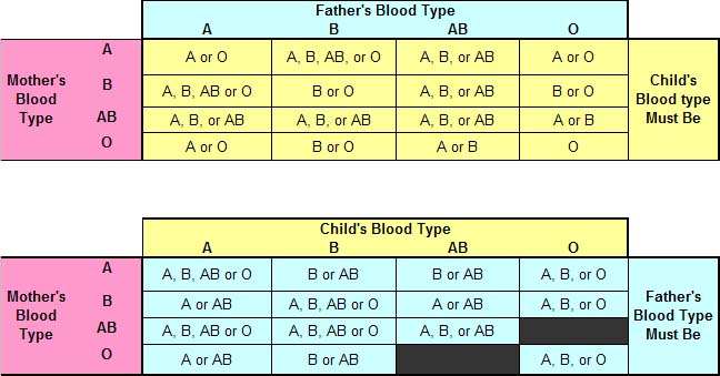
Mechanism of inheritance
of blood group
A and B are dominant antigen and O
is recessive antigen. Depending on the genotype of parents the group of
offspring can be predicted. Examples;
Rh factor- Rh factor was discovered by
K.Landsteiner and Wiener in 1940 in RBCs of Rhesus monkey (Macca rhesus).This antigen is found on the surface of RBC. Most of
85% of people have Rh factor. The presence of Rh factor in blood of a person is
called Rh +Ve and absence is called Rh –Ve. Rh +Ve and Rh –Ve are incompatible
and cannot be mixed. It is great importance in two conditions:
1. During
transfusion of blood 2. During pregnancy
Rh incompatibility
Rh factor is very
important as it is inheritable. When Rh+ man marries a Rh- woman there is a
chance that some of their children will be Rh+. During pregnancy, fragments of
Rh+ RBC of the foetus may enter blood of mother. It act as antigen and mother body produce antibodies against
the Rh+.The bodies passes from placenta to the foetus and destroy foetal red
cell. It happens in second pregnancy because in first pregnancy the antibody
will form but not enough to destroy RBC of foetus. The baby born to be
premature, anaemic and jaundice. The condition is called erythroblastosis foetalis.
About one in every
10 marriages is between Rh+men and Rh- women, but only about one in 40 of these
marriages is affected by the Rh- incompatibility.
ReplyDeleteBest ias coaching in bangalore
.www.globalias.in
Diabetes called a group of metabolic disorder. Cause it can occur many serious types of complication such as kidney disease, cardiovascular, stroke foot ulcer. Healing Diabetes Treatment has provided by Stem Cells.
ReplyDelete